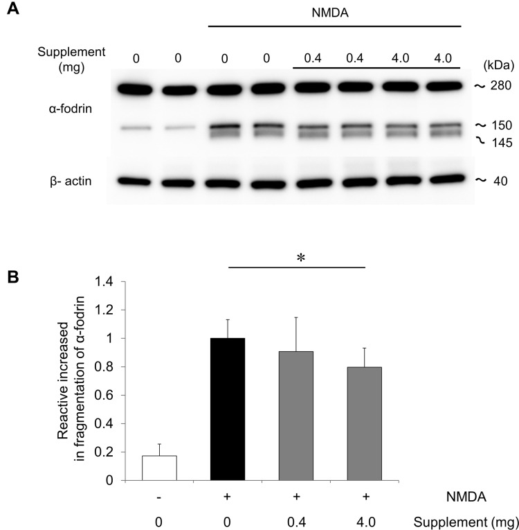
Figure 2.
Oral supplementation reduced the cleavage of α-fodrin in the retina after NMDA injury. (A) Immunoblot analysis of α-fodrin in retinas without supplementation or with a low- or high-dose supplement 6 hours after NMDA injury. Representative immunoreaction image with anti-α-fodrin showing intact α-fodrin (280 kDa) and calpain-cleaved fragmented α-fodrin (145 and 150 kDa). β-actin was used as an internal control. (B) The relative density of the cleaved-fodrin immunoreactive band. Relative density was based on the cleaved-fodrin immunoreactive band 6 hours after NMDA injection. Data represent mean ± SD (each group: n = 6). *p <0.05.

