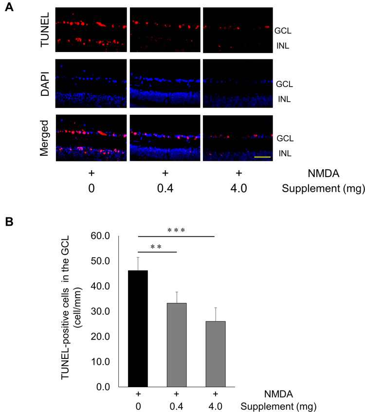
Figure 3.
Decrease in TUNEL-positive cells after NMDA injury and supplementation. (A) Representative overlay photographs of retinal sections in mice with or without supplementation 24 hrs after NMDA injection. Red: TUNEL assay; blue: DAPI nuclear staining. Scale bar: 100 µm. (B) Histograms showing the TUNEL-positive cell count in the GCL of mice (non-supplementation group: n = 7, other groups: n = 8). Data represent mean ± SD, **P < 0.01, ***P < 0.001.
Abbreviations: GCL, ganglion cell layer; INL, inner nuclear layer.

