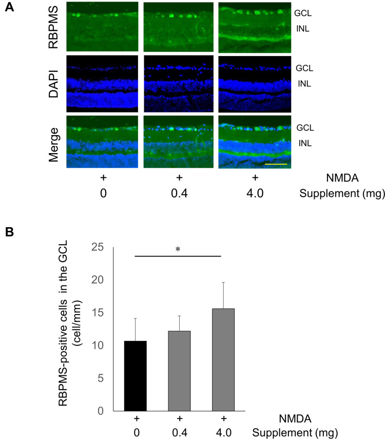
Figure 4.
Increase in RBPMS-positive cells after NMDA injury and supplementation. (A) Representative images of RBPMS-positive RGCs 24 hours after the intravitreal injection of NMDA without supplementation or with a low- or high-dose supplement. GCL, ganglion cell layer; INL, inner nuclear layer. Scale bar: 100 µm. (B) Histogram showing the average number of RBPMS-positive cells in each group. Data represent mean ± SD (PBS, n = 8; low-dose supplement, n = 6; high-dose supplement, n = 8). *p <0.05.

