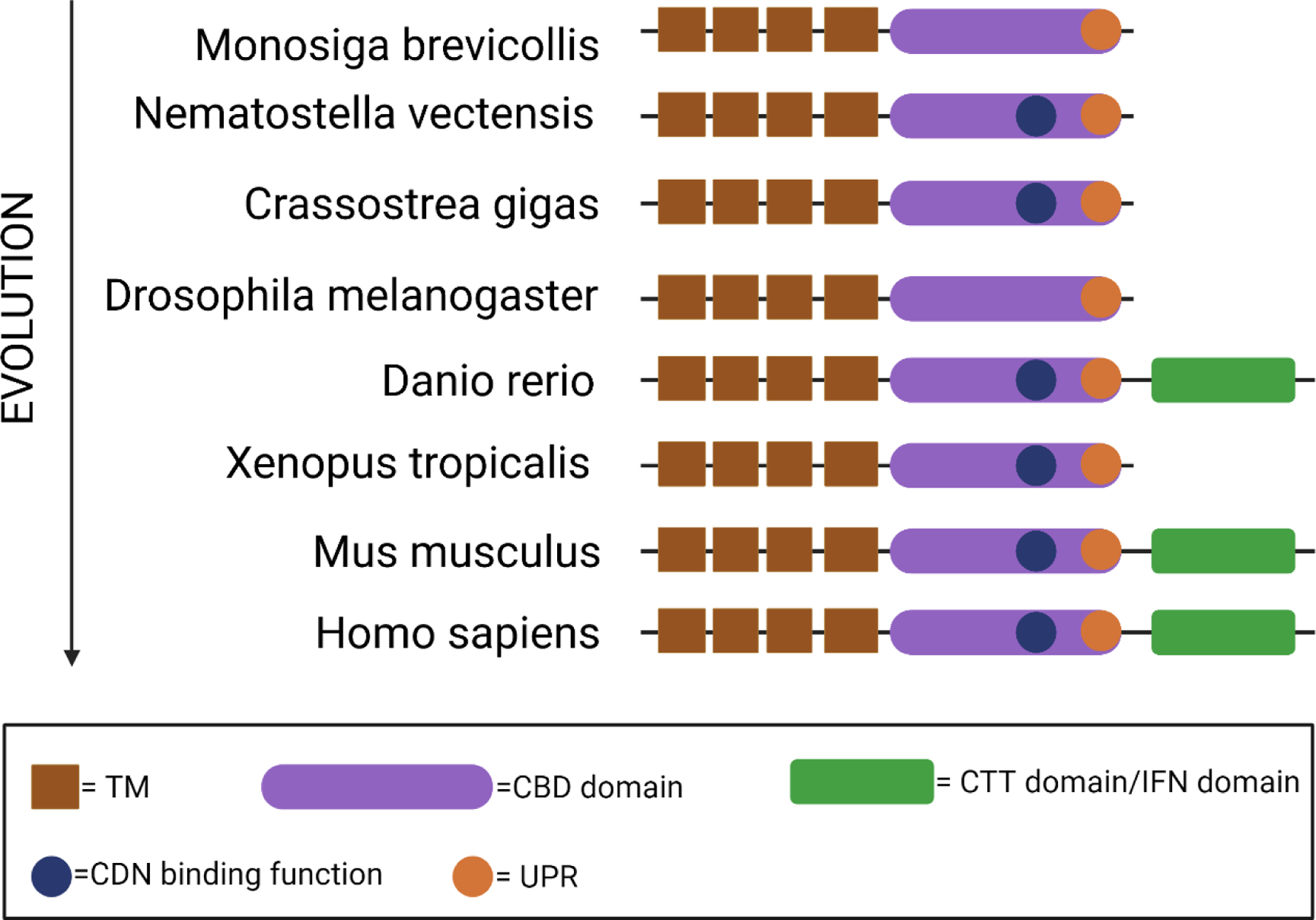
Figure 1. The evolution of STING protein.

The evolution of STING protein homologs from different species [35, 36] is illustrated by their evolutionary hierarchy. Conserved structure domains and motifs are represented with a diagram on the right. TM, transmembrane; CBD: cyclic dinucleotide binding domain; CDN, cyclic dinucleotide, CTT, C-terminal domain; UPR, unfolded protein response.
