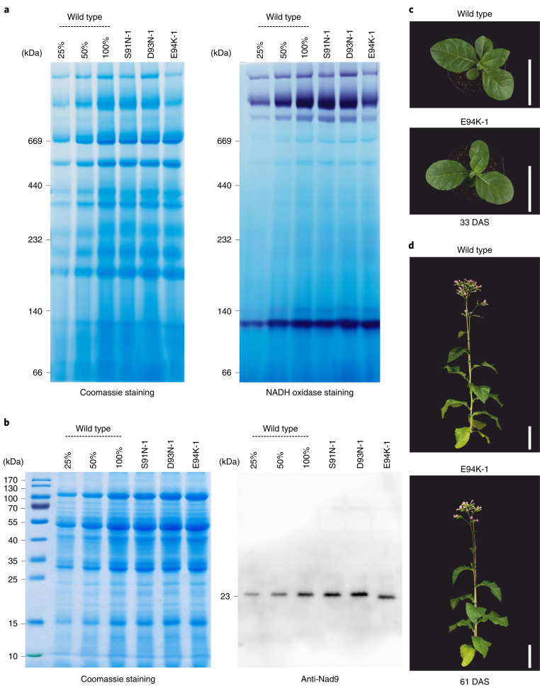
Fig. 4. Biochemical characterization and phenotypes of mitochondrial nad9 mutants.
a, Analysis of mitochondrial protein complexes and NADH oxidase (complex I) staining in selected nad9 mutants by blue-native polyacrylamide gel electrophoresis (BN–PAGE). Protein sizes are given in kDa. A dilution series of the wild-type sample (25%, 50% and 100%) was loaded to allow for semiquantitative assessment of protein accumulation. A technical replicate of the BN–PAGE (stained with Coomassie) yielded similar results; the NADH oxidase (complex I) staining was performed once. b, Accumulation and electrophoretic mobility of Nad9 proteins in nad9 mutants assessed by SDS–PAGE. Note the faster migration of the E94K variant of Nad9. The Coomassie-stained gel is shown to confirm equal loading. The SDS–PAGE (including Coomassie staining) was done seven times and the anti-Nad9 western blot five times (technical replicates), with similar results (see also Extended Data Fig. 9). c, Plant phenotypes. Plants were grown on soil and cultivated in long-day conditions in the greenhouse. A wild-type plant and an E94K-1 mutant plant (harbouring a G280A mutation in nad9) are shown. The photographs were taken 33 days after sowing (DAS). Scale bars, 10 cm. d, Plants photographed 61 DAS. Scale bars, 20 cm.

