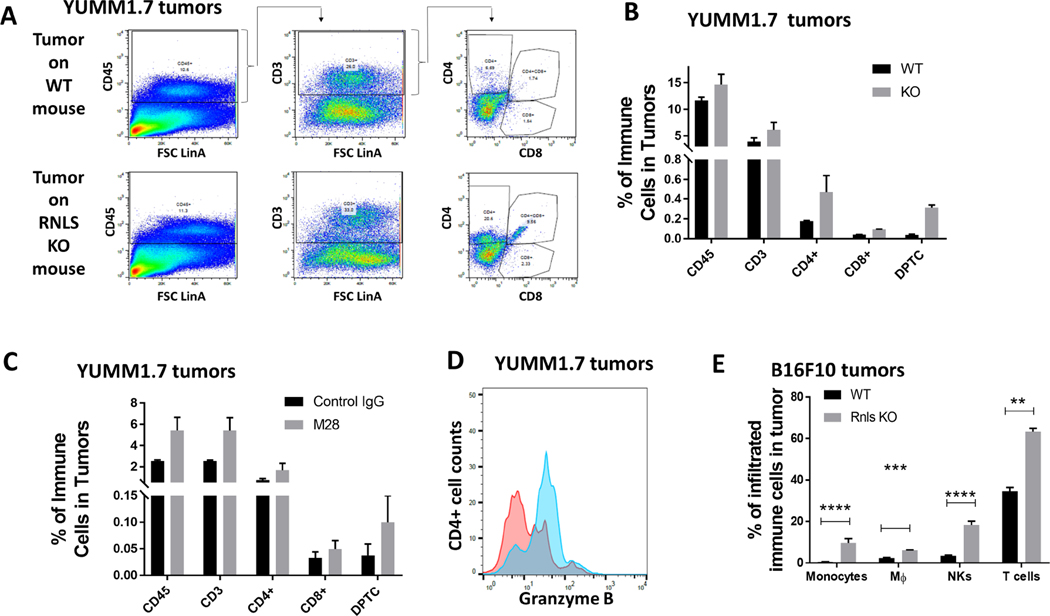
Figure 3.
Protein level changes induced by RNLS knock-out or inhibition confirm changes seen with scRNA-seq: (A-B) Flow cytometry of T cells in YUMM1.7 tumors implanted on WT or RNLS- KO mice showing increased CD4 and CD8 content. (C) m28-RNLS treatment results in similar increases in T cell content, and CD4 T cell activation as shown by Granzyme B increases (D). Increases in monocytes, macrophages, NK and T cells were confirmed in B16F10 tumors at the protein level (E).

