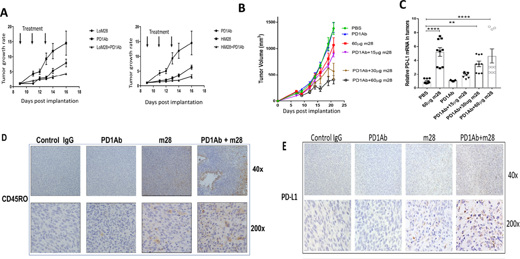
Figure 5.
Inhibition or elimination of RNLS in combination with anti-PD-1 is superior to either modality alone in two murine melanoma models. (A) Mice bearing B16F10 tumors treated with two doses of m28-RNLS (60ug and 120ug) alone or with anti-PD-1. Combination therapy was superior to either drug alone, N=5 per cohort. (B) Confirmation studies done in mice bearing YUMM1.7 tumors treated with 15 μg, 30 μg or 60 μg m28-RNLS with or without anti-PD-1, demonstrating superior activity of the combination compared to either drug alone (N=5 per cohort). (C) mRNA levels of PD-L1 are higher in tumors from mice treated with m28-RNLS, suggesting a potential mechanism for sensitization of tumors to anti-PD-1 by m28-RNLS. (D-E) Immunohistochemical studies demonstrating increased memory T cell content and increased tumor PD-L1 expression with combined therapy compared to either drug alone. Control IgG treated tumors had a mean of 1.6±0.84 CD45RO positive cells per high power field (400X), anti- PD-1 treated tumors had 3.4±0.9, m28 tumors had 22±1.4, and tumors treated with the combination had 31±3 CD45RO positive cells, markedly more than control or anti-PD-1 alone (P<0.0001, by Tukey’s test for multiple comparisons). We found a mean of 2.2±0.8 PD-L1 positive cells per high power field (400X) in control IgG treated tumors, 2.3±0.5 in anti-PD-1 treated tumors, 19.9±3.4 in m28 treated tumors and tumors treated with the combination had 43.3±3.6 PD-L1 positive cells, markedly more than control or either drug alone (P<0.0001, by Tukey’s test for multiple comparisons).

