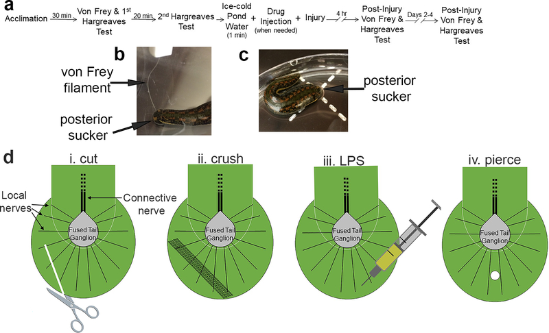
Figure 1:
Experimental methods for studying injury-induced sensitization in Hirudo. (a) Timeline for experimental protocol. (b) Example of application of a von Frey filament to the posterior sucker of Hirudo. (c) Example of Hirudo in a petri dish set over the Hargreaves apparatus (indicated by the dashed plus sign). (d) Graphics illustrating basic anatomy of the posterior sucker and the four injury-inducing protocols in the posterior sucker. The sucker is innervated by 14 local nerves (only three are labeled) that radiate from the fused tail ganglion. The tail ganglion is in the portion of the leech body that terminates just before the posterior sucker and this ganglion is connected to anterior segmental ganglia via the connective nerve (Muller et al. 1981). The terminal portion of the leech body also includes musculature and elements of the vascular system and rectum (not shown) and this is the region where the THL injections were carried out. (i) Cut protocol using surgical scissors. (ii) Crush protocol using a hemostat. (iii) LPS injection protocol. (iv) Piercing injury protocol using a T-pin.

