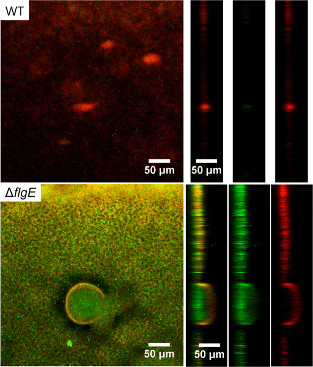
Fig. 6. Confocal laser scanning microscopy observation of MPAO1 WT and ΔflgE biofilms after gentamicin treatment.

Forty hours-old biofilms were exposed to M9 medium supplemented with 12 µg/mL of gentamicin for 24 h. SYTO9 and PI were used to stain living bacteria in green and membrane damaged bacteria in red, respectively. Orthogonal views of the mushroom-like biofilm are shown from left to right with an overlay of SYTO9 and PI, SYTO9 alone and PI fluorescence alone.
