Figure 2.
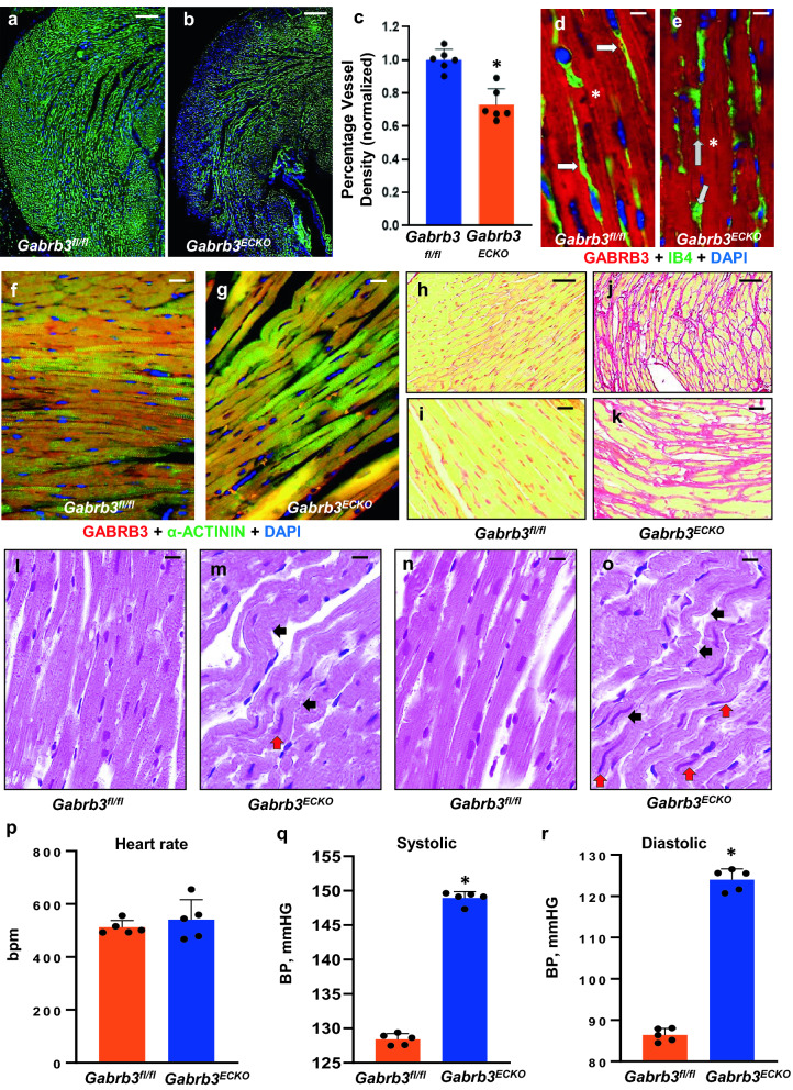
Gabrb3ECKO mice have abnormal cardiac pathology and hypertension. (a–c) Isolectin B4-labeled vessels were significantly reduced in Gabrb3ECKO hearts, when compared to floxed controls (a, b). Vessel quantification depicted in (c); Data represent mean ± SD (n = 6, *P < 0.05; Student's t-test). (d, e) Co-labeling with Isolectin B4 (red) and GABRB3 (green) revealed expression of GABRB3 in Gabrb3fl/fl vessels only (merged in yellow, white arrows, (d) and its lack thereof in Gabrb3ECKO vessels (gray arrows, e). Prominent expression of GABRB3 was observed in cardiomyocytes in both groups (asterisk). (f, g) Validation of GABRB3 expression in cardiomyocytes by co-labeling with GABRB3 and α-ACTININ in Gabrb3fl/fl and Gabrb3ECKO mice. (h–k) Low (h, j) and high (i, k) magnification images of normal picrosirius red staining in Gabrb3fl/fl cardiac tissue (h, i), and an increase in collagen deposition, observed in Gabrb3ECKO cardiac tissue (j, k). (l-o) H & E staining shows wavy myocardial fibers (black arrows, m, o) with long ‘worm-like’ nuclei (red arrows, m, o) in Gabrb3fl/fl cardiac tissue, which is not seen in controls (l, n). (p) No changes in heart rate were observed in Gabrb3fl/fl and Gabrb3ECKO mice. (q, r) A significant increase in systolic and diastolic blood pressure was observed in Gabrb3ECKO mice versus Gabrb3fl/fl mice. Data represent mean ± SD (n = 5, *P < 0.05, Student's t-test). Scale bars: (a), 100 μm (applies to b, h, j); d, 50 μm (applies to e, f, g, i, k, l-o).

