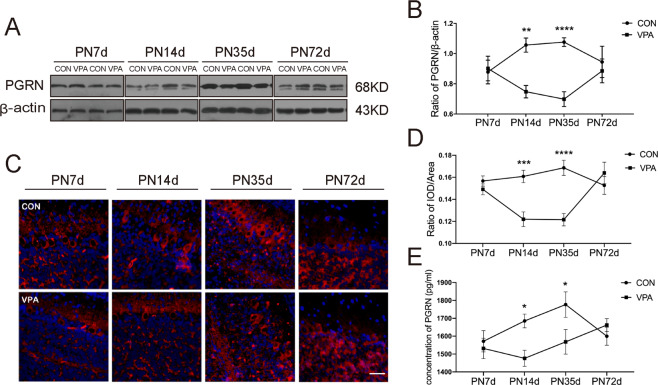
Fig. 1. Prenatal exposure to VPA caused the alterations of PGRN temporal expression in the cerebellum.
Representative blots (A) and quantification (B) showed the expression of PGRN at four different time points. Representative images (bar = 100 μm) (C) and quantification (D) of immunofluorescence staining described that the expression of PGRN is in the same trend as Western blotting. E PGRN ELISA protein quantitation of cerebellar tissue. Data are expressed as mean ± SEM. Two-way ANOVA followed by Sidak post hoc. (*P < 0.05, **P < 0.01, ***P < 0.001, ****P < 0.0001, sample sizes (n): n = 5/group for Western blotting and immunofluorescence staining. n = 4/group for ELISA).

