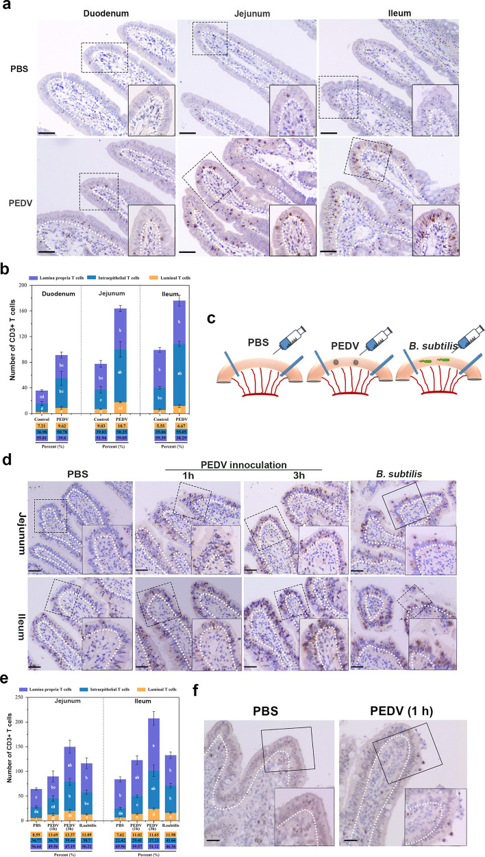
Fig. 2. Influence of porcine epidemic diarrhea virus (PEDV) infection on porcine intestinal intraepithelial lymphocytes (IELs).
a Pigs (n = 3) from mock or PEDV challenge groups were euthanatized at 48 hpi, and small intestinal tissues were fixed and subjected to IHC analysis. Representative image of IELs distribution in the small intestine of PBS-inoculated or PEDV-inoculated weaned piglets (one-month-old). Scale bars, 20 μm. b Quantitative analysis of IELs showed in (a) was performed, in which 20 random intestinal villi were counted. The frequency of total T cells in each area (%) were also presented. c Schematic of the experimental setting of the intestinal ligated loop model. Anesthetized piglets (one-month-old, n = 3) were subjected to intestinal ligation followed by injection with PBS, PEDV, and Bacillus subtilis, and were euthanatized at 3-hours post-treatment. d Immunohistochemistry (IHC) analysis demonstrated the distribution pattern of IELs influenced by PEDV or B. subtilis in the ligated loop of the terminal jejunum. Scale bars, 20 μm. e Quantification of IELs (d) in 20 random intestinal villi. The frequency of total T cells in each area (%) was also calculated. f The proliferative activity of intraepithelial and transepithelial IELs in the ligated loop was detected via proliferating cell nuclear antigen (PCNA) staining. The scale bar represents 50 μm. All data shown are the mean results ± SD from three independent experiments. Statistical significance was obtained using one-way ANOVA. NS no significance, *P < 0.05, **P < 0.01. The differences are indicated by different letters. Letters above the graphs indicate statistical significance in which treatments with a letter in common are not significantly different from each other.

