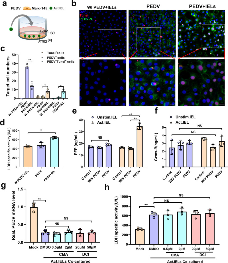
Fig. 7. The antiviral activity of IELs was independent from their cytotoxic effects.
a The design for detecting the cytotoxicity of IELs. The virus pre-activated IELs were co-cultured with porcine epidemic diarrhea virus (PEDV)-infected epithelial cells for 24 h. b TUNEL staining was used to determine the apoptosis rate of PEDV-infected or whole inactivated PEDV-treated epithelial cells. c The number of TUNEL+, PEDV+, and TUNEL+PEDV+ cells in each group was calculated and shown in a bar graph, which was produced from five random fields of view for each of three individual sections. d The released lactate dehydrogenase (LDH) activity assay was also used to evaluate the cytotoxicity of IELs in different groups. e, f Levels of perforin (e) and granzyme B (f) release in the culture supernatants of different groups were measured via ELISA. g, h The pre-reactivated IELs were treated with CMA at two concentrations (1 and 0.5 μM) or DCI at two concentrations (20 and 50 μM) for 3 h, and their antiviral activity were further detected by co-culturing with virus-infected epithelial cells. Both of the inhibitors were also maintained for the whole duration of the co-culturing process. The intracellular viral RNA expression (g) and extracellular LDH activity (h) in each experimental group were determined. All data are the mean ± SD and comparisons were performed using one-way ANOVA. *P < 0.05, **P < 0.01. The results are from at least three different experiments.

