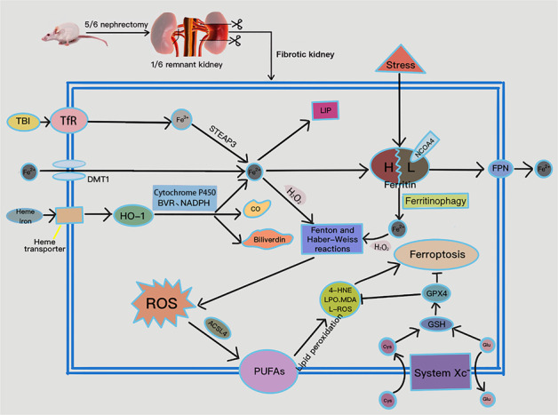
Fig. 7. Disordered iron metabolism and ferroptosis occurred in 5/6 nephrectomy-induced CKD rats.

DMT1 and TfR expression were down-regulated, while HO-1 was up-regulated; the latter catabolizes heme iron to ferrous iron, CO, and biliverdin, with the assistance of cytochrome P450, biliverdin reductase (BVR), and NADPH. TfR mediates the import of transferrin-bound iron (TBI) into the cell and reduces ferric iron to ferrous iron via six-transmembrane epithelial antigen of prostate 3 (STEAP3). Regarding iron export, FPN down-regulation blocks iron efflux, leading to elevated intracellular iron concentration. A small amount of intracellular ferrous iron is stored in the labile iron pool (LIP) to maintain physiological metabolism, while the remainder is endogenously chelated by ferritin. Excess intracellular catalytic iron reacts with hydrogen peroxide to initiate the Fenton and Haber-Weiss reactions, leading to a further increase in ROS and attacks on polyunsaturated fatty acids (PUFAs) in the cytomembrane, initiating lipid peroxidation and ferroptosis. In addition, down-regulation of SLC7A11 results in reduced synthesis of GSH from Cys, decreased GPX4 activity, and weakened anti-ferroptotic responses. CO carbon monoxide, Glu glutamate.
