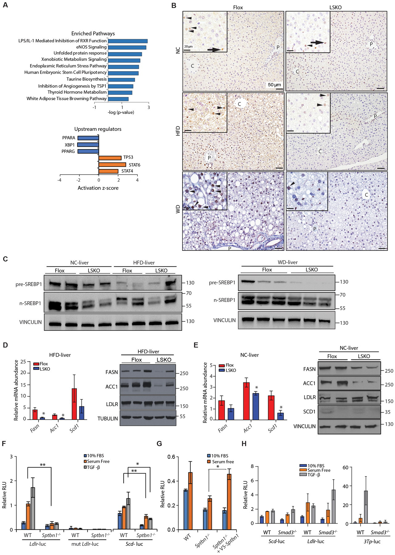
Fig. 2. SREBP1 signaling is reduced in LSKO mouse livers in both the normal chow and HFD conditions.

(A) Bioinformatic analysis by Ingenuity Pathway Analysis of differentially regulated genes in livers from LSKO mice compared to Flox mice after 16 weeks of HFD. Differentially regulated genes were determined from RNA-seq data from 5 mice in each group. Upper: Pathway enrichment analysis. Lower: Upstream regulator analysis for regulators with activation (z score ≥2) or inhibition (z score ≤ 2), showing those with p < 0.05.
(B) Immunohistochemistry of SREBP1 in liver sections from Flox and LSKO mice after 16 weeks of feeding with NC, HFD, or WD using anti-SREBP1 from Abcam (ab191857). Black arrowheads indicate hepatocytes nuclear localization, black arrow indicates labeling in non-hepatocytes. C-central vein; P-portal vein.
(C) Analysis of livers from Flox and LSKO mice fed NC, HFD or WD by Western blot for full-length SREBP1 and the active cleaved forms (n-SREBP1) using anti-SREBP1 from Abcam (ab191857). Each lane represents samples from a single mouse (n = 2 or 3).
(D) Relative mRNA and protein abundance for products of SREBP1 target genes from livers of LSKO and Flox mice after 12 to 16 weeks of feeding with HFD. RNA data are normalized to 18S (n = 6 – 7 for each group). Western blot data show results from individual mice.
(E) Relative mRNA and protein abundance for products of SREBP1 target genes (Fasn, Acc1, Scd1) and a SREBP2 target gene (Ldlr) from livers of LSKO and Flox mice fed normal chow (NC). RNA data are normalized to 18S (n = 3 – 5 for each group for). Western blot data show results from individual mice.
(F) SRE-driven luciferase activity from the LDLR-luc, mutant LDLR-luc, and SCD-luc reporters in WT and Sptbn1−/− MEF cells after 24 hours in medium with 10% fetal bovine serum (FBS) or serum-free medium with or without TGF-β1 (200 pM) for 24 hours.
(G) SRE-driven luciferase activity from the LDLR-luc reporter in WT and Sptbn1−/− MEF cells expressing empty vector or SPTBN1 and cultured for 24 hours in 10% FBS or serum-free medium.
(H) Luciferase activity from the SCD-luc and LDLR-luc reporters (SREBP-responsive) and 3TP-luc reporter (SMAD3-responsive) in WT and Smad3−/− MEF cells after 24 hours in medium with 10% fetal bovine serum (FBS) or serum-free medium with or without TGF-β1 (200 pM) for 24 hours.
Quantitative data shown as mean ± SEM in panels D to H are representative performed in triplicate or summarized from 2–3 independent experiments performed in triplicate. Statistical significance was determined by 2-sided t-test or one-way ANOVA (*, p < 0.05; **, p < 0.005).
