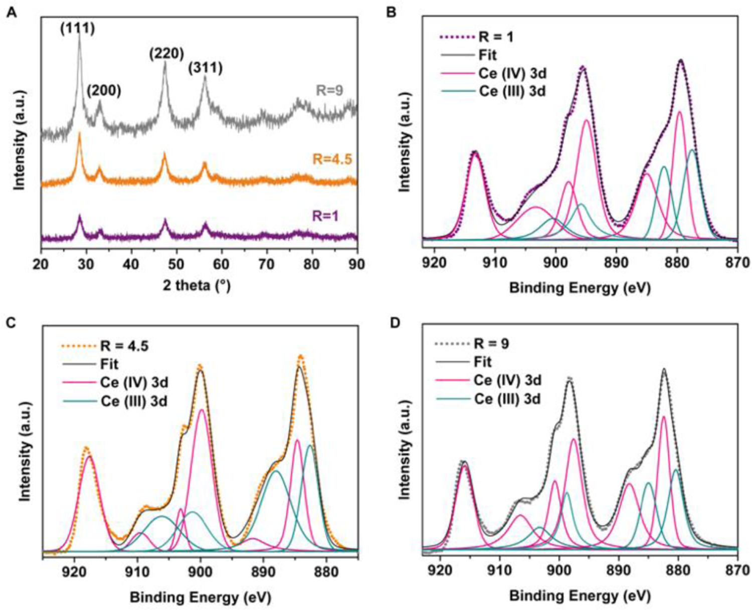
Fig. 2.

(A) XRD patterns of the CeO2-x nanorods. (B-D) XPS high-resolution scan of Ce(3d) in the CeO2-x nanorods with R ratios of B) R=1, C) R=4.5; and C) R=9, and the peak deconvolution results. Dots indicate raw data; black solid lines correspond to the fitted curves based on peak deconvolution.
