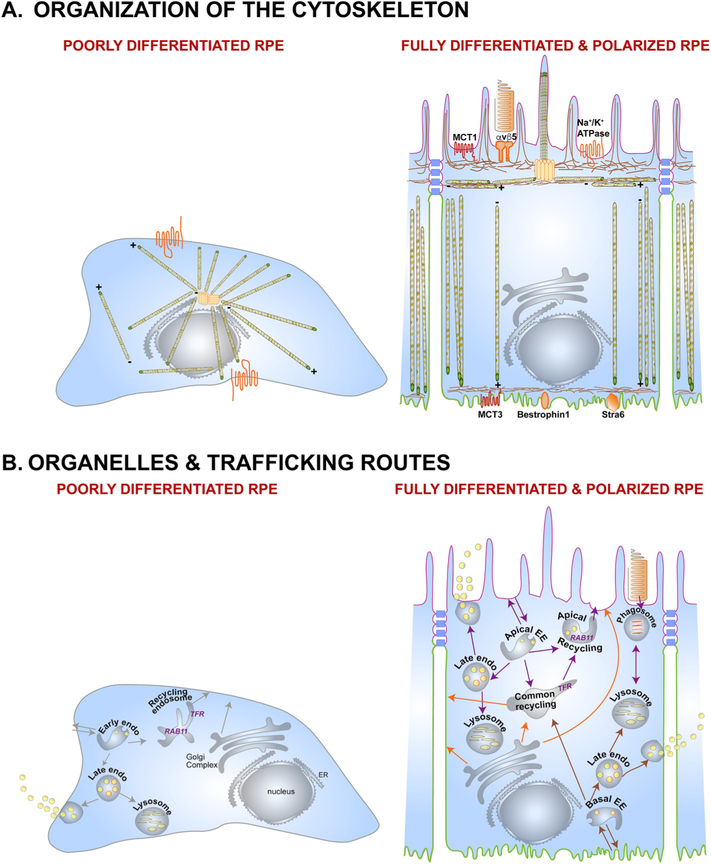
Fig. 2.
The polarized phenotype of the RPE. (A) Organization of the cytoskeleton in the RPE. In a non-polarized RPE cell (left), microtubules originate at the microtubule organizing center (MTOC), with the plus ends oriented towards the cell periphery. This poorly differentiated RPE cell shows non-polarized distribution of membrane proteins like the Na+/K+-ATPase (orange). A fully differentiated RPE cell (right) has well-defined apical (pink) and basolateral (green) membrane domains demarcated by tight junctions (blue). Microtubules are organized vertically with the plus ends oriented towards the basal surface. A lateral microtubule network and the cortical actin network are also found below the microvilli. This precisely organized cytoskeletal network provides structural support for the RPE and ensures accurate localization of apical (e.g., αvβ5, MCT1, Na+/K+-ATPase) and basolateral (e.g., MCT3, Bestrophin1, Stra6) membrane proteins. (B) Organelles and trafficking routes in the RPE. Polarized RPE have distinct biosynthetic (orange arrows), apical (purple arrows), and basolateral (brown arrows) trafficking routes that transport specific cargo destined for specific membrane domains, or for secretion into the apical or basolateral extracellular space. A key feature of polarized epithelia like the RPE is the apical recycling endosome, which is identified by the presence of RAB11, and distinct from the common recycling endosome, which has the transferrin receptor. In a non-polarized cell, RAB11 is found along with the transferrin receptor in the common recycling endosome. EE, early endosome; ER, endoplasmic reticulum.

