Fig. 5.
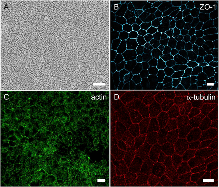
Differentiated cultures of ARPE-19 cells. (A) Brightfield micrograph showing cobblestone morphology of ARPE-19 cells cultured on plastic for 2 weeks. (B) Immunofluorescence micrograph of zonula occludens (ZO)-1 localized to the apical junctions of ARPE-19 cells cultured on a laminin-coated Transwell filter for 2 weeks. (C) Phalloidin labeling in ARPE-19 cells cultured on a laminin-coated Transwell filter for 3 weeks shows cortical arrangement of the actin filaments. (D) Micrograph of α-tubulin immunolabeling demonstrating vertical microtubule arrangement in ARPE-19 cells cultured on a laminin-coated Transwell filter for 3 weeks. The cells depicted in all four panels were differentiated using the protocol described in (Hazim et al., 2019). Scale bar in A = 100 μm, (B–D) = 10 μm.

