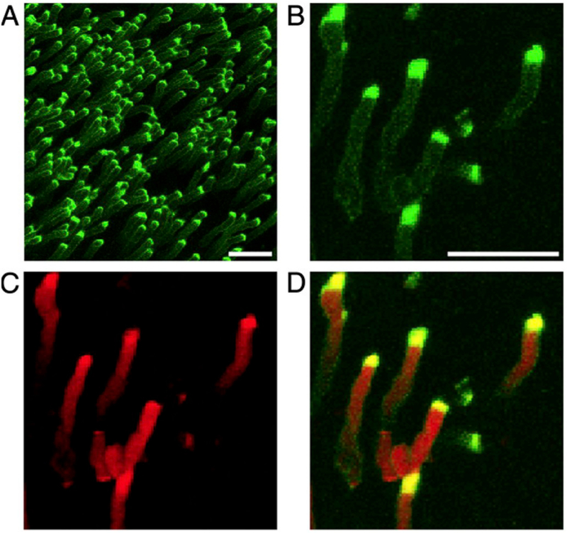
Fig. 8.
Phosphatidylserine (PS) exposure at the tips of photoreceptor outer segments (OS). Wild type mouse retina dissected live at light onset are labeled and imaged with (A) a polarity-sensitive indicator of viability and apoptosis (pSIVA; green), which specifically binds to PS residues exposed to the extracellular space. (B–D) Higher magnification images show co-staining of (B) pSIVA and the (C) CellMask membrane stain (red), and (D) an overlay of both. Scale bar in A = 10 μm, B for B–D = 5 μm. Originally published in the Proceedings of the National Academy of Sciences (Ruggiero et al., 2012); permission granted by PNAS.

