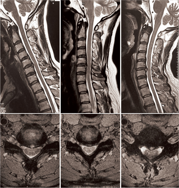
Fig. 3.
Dynamic MR images of a patient with improved DSSEPs at extension and deteriorated DSSEPs at flexion. Panels from left to right show cervical flexion, neutral and extension positions. Upon neutral positioning, this patient had a straight cervical alignment and a single protruding C5/6 segment. The Mühle stenosis grade of this patient was Grade 1. Upon flexion, the spinal cord was longitudinally stretched and draped backward. The cerebral fluid in front of the spinal cord was narrower in the axial image. Upon extension, although the diameter of his cervical canal did not change significantly, the spinal cord was longitudinally relaxed and draped backward and, therefore, ameliorated anterior compression to some extent. The cerebral fluid in front of the spinal cord was wider in the axial image captured in the extension position

