
FIG 3.
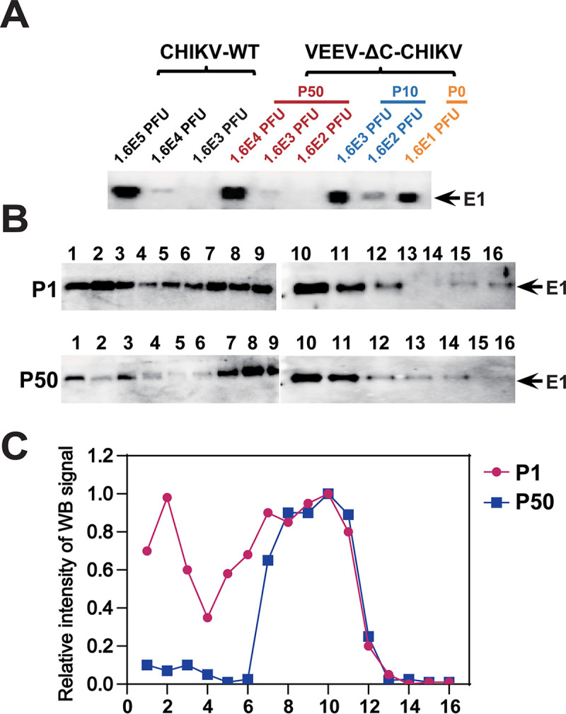
The passaged VEEV-ΔC-CHIKV produces more infectious virus particles. (A) Western blotting of CHIKV E1 expression of different viral loads of CHIKV-WT and VEEV-ΔC-CHIKV at P0, P10, and P50 using CHIKV E1 polyclonal antibodies. (B) Western blotting of sucrose density gradient fractionations of VEEV-ΔC-CHIKV at P0 and P50 produced in Vero cells. 105 PFU P0 and 107 PFU P50 viruses were separated on 20%–60% linear sucrose density gradients. Sixteen fractions were harvested from the top (Fraction 1) to the bottom (Fraction 16) of the gradient. Each fraction from P0 and P50 VEEV-CHIKV was subjected to Western blotting assay using E1-specific antibody. (C) Analysis of the intensity of viral E1 protein bands in each fraction using ImageJ software.