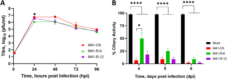
FIG 2.
The replication kinetics of M41-R are comparable to those of M41-CK in vitro and in ex vivo TOCs. (A) Primary CK cells were inoculated with 105 PFU of either rIBV M41-R-6, M41-R-12, or M41-CK. Supernatants were harvested at 24-h intervals and quantities of infectious progeny virus present were determined by plaque assay in CK cells. Each point represents the mean of three independent experiments with error bars representing standard error of the mean (SEM). (B) Ex vivo TOCs, prepared from 19-day-old SPF embryos, were inoculated in replicates of 11 with 104 PFU of either rIBV M41-R-6, M41-R-12, M41-CK, or medium for mock infection. Ciliary activities were assessed by light microscopy at regular intervals and the mean activities of 11 replicates were calculated. Error bars represent SEM. (A and B) Statistical differences were analyzed using a two-way ANOVA with Tukey analysis for multiple comparisons and are indicated by * (P < 0.05) and **** (P < 0.0001). For clarity, in panel A, * denotes that rIBV M41-R-6 is lower than both rIBV M41-R-12 and M41-CK at 24 hpi.

