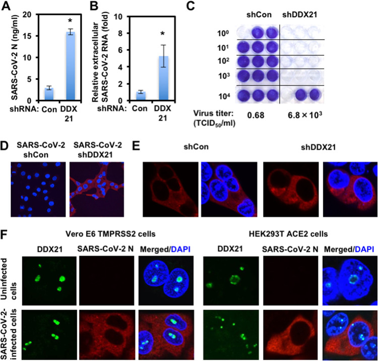
FIG 2.
Characterization of antiviral effect of DDX21. (A) DDX21 suppresses SARS-CoV-2 production. The levels of extracellular SARS-CoV-2 N protein in the culture supernatants from the DDX21 knockdown HEK293T ACE2 cells 72 h after inoculation of SARS-CoV-2 at an MOI of 0.5 were determined by ELISA. Experiments were done in triplicate, and columns show the mean SARS-CoV-2 N protein levels. *, P < 0.05 compared to control cells. (B) DDX21 inhibits the level of extracellular SARS-CoV-2 RNA. The levels of extracellular SARS-CoV-2 RNA in the culture supernatants from the DDX21 knockdown HEK293T ACE2 cells 72 h after inoculation of SARS-CoV-2 at an MOI of 0.5 were monitored by real-time LightCycler RT-PCR. Results from three independent experiments are shown. The level of SARS-CoV-2 RNA in DDX21 knockdown cells was calculated relative to the level in HEK293T ACE2 cells transduced with a control lentiviral vector (Con). *, P < 0.05 compared to control cells. (C) The virus titer of SARS-CoV-2 in the culture supernatants from the DDX21 knockdown HEK293T ACE2 cells 72 h after inoculation of SARS-CoV-2. Naive Vero E6 TMPRSS2 cells were seeded in 24-well plates at 5 × 10⁴ cells per well and then infected the next day with the indicated serial 10-fold dilutions of culture supernatants. The cells were stained with 0.6% Coomassie brilliant blue in 50% methanol and 10% acetate at 72 h postinfection were monitored for cytopathic effect. The virus titer was determined as TCID50/mL. (D) The infectivity of SARS-CoV-2 in the culture supernatants from the control or DDX21 knockdown HEK293T ACE2 cells 72 h after inoculation of SARS-CoV-2 was compared by immunofluorescence. Naive Vero E6 TMPRSS2 cells were plated on Lab-Tek 2-well chamber slides at 2 × 10⁴ cells per well. The next day, 1 μL of culture supernatants of SARS-CoV-2-infected control or DDX21 knockdown HEK293T ACE2 cells was inoculated. The cells were fixed at 24 h postinfection and stained with anti-SARS-CoV-2 nucleocapsid (ab273434 [6H3]). Cells were then stained with donkey anti-mouse IgG (H+L) Alexa Fluor 594-conjugated secondary antibody. Images were visualized using confocal laser scanning microscopy. Nuclei were stained with DAPI (blue). (E) Subcellular localization of SARS-CoV-2 N protein in control or DDX21 knockdown HEK293T ACE2 cells 24 h after inoculation of SARS-CoV-2. The cells were stained with anti-SARS-CoV-2 nucleocapsid. (F) Subcellular localization of endogenous DDX21 and SARS-CoV-2 N protein in Vero E6 TMPRSS2 or HEK293T ACE2 cells 24 h after inoculation of SARS-CoV-2. The cells were stained with anti-SARS-CoV-2 nucleocapsid and anti-DDX21 (A300-627A) antibodies. Cells were then stained with donkey anti-rabbit IgG (H+L) Alexa Fluor 488-conjugated secondary antibody and donkey anti-mouse IgG (H+L) Alexa Fluor 594-conjugated secondary antibody. The two-color overlay images are also exhibited (Merged).

