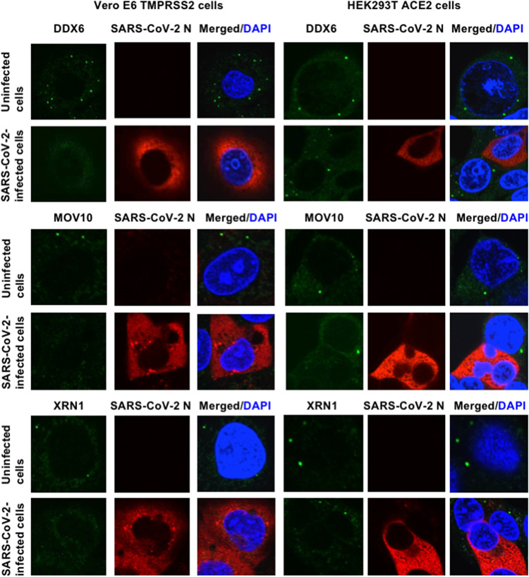
FIG 4.
SARS-CoV-2 disrupts P-body formation. Uninfected Vero E6 TMPRSS2 or HEK293T ACE2 cells and their SARS-CoV-2-infected cells at 24 h postinfection were stained with anti-SARS-CoV-2 nucleocapsid (ab273434 [6H3]) and anti-DDX6 (A300-460A) antibodies. The cells were also stained with anti-SARS-CoV-2 nucleocapsid and either anti-Xrn1 (A300-443A) or anti-MOV10 (A301-571A) antibodies. Cells were then stained with donkey anti-rabbit IgG (H+L) Alexa Fluor 488-conjugated secondary antibody and donkey anti-mouse IgG (H+L) Alexa Fluor 594-conjugated secondary antibody. Images were visualized using confocal laser scanning microscopy. The two-color overlay images are also exhibited (Merged). Nuclei were stained with DAPI (blue).

