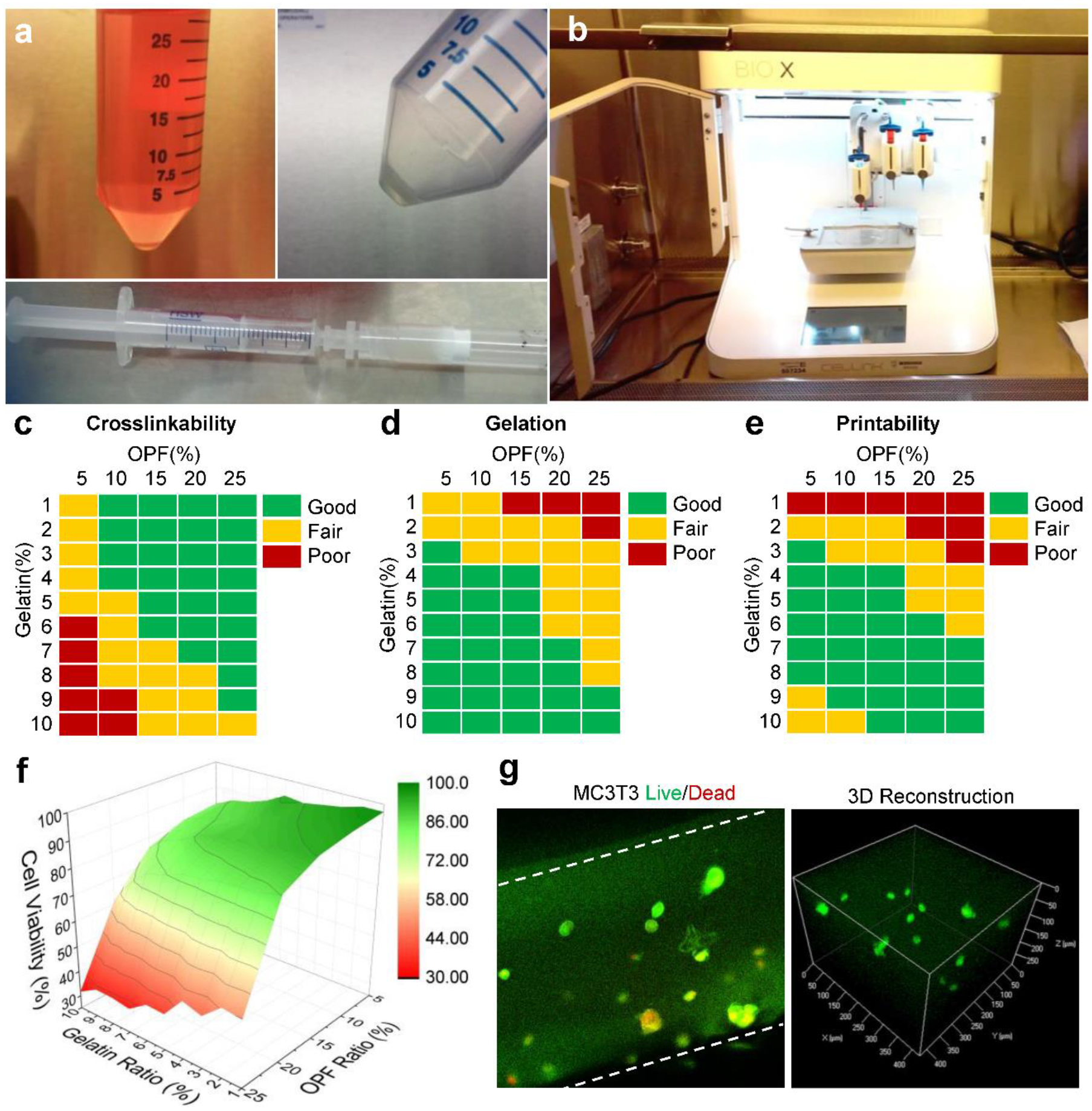
Fig. 3.

a) The collection of MC3T3 pre-osteoblast cells from the culture medium and mix with liquidized gels to obtain the bioink. b) Photographs of the bioprinting apparatus set-up using a CELLINK Bio-X printer in a sterilized cell culture hood. The c) crosslinking ability, d) gelation performance and e) printability of bioinks with varied OPF and gelatin concentrations. f) Cell viabilities of bioinks with varied OPF and gelatin concentrations. g) Confocal imaging of LIVE/DEAD stained cells encapsulated within the bioprinted scaffolds: live cells (green) and dead cells (red). Cell density: 1 × 10⁶ cells/mL medium.
