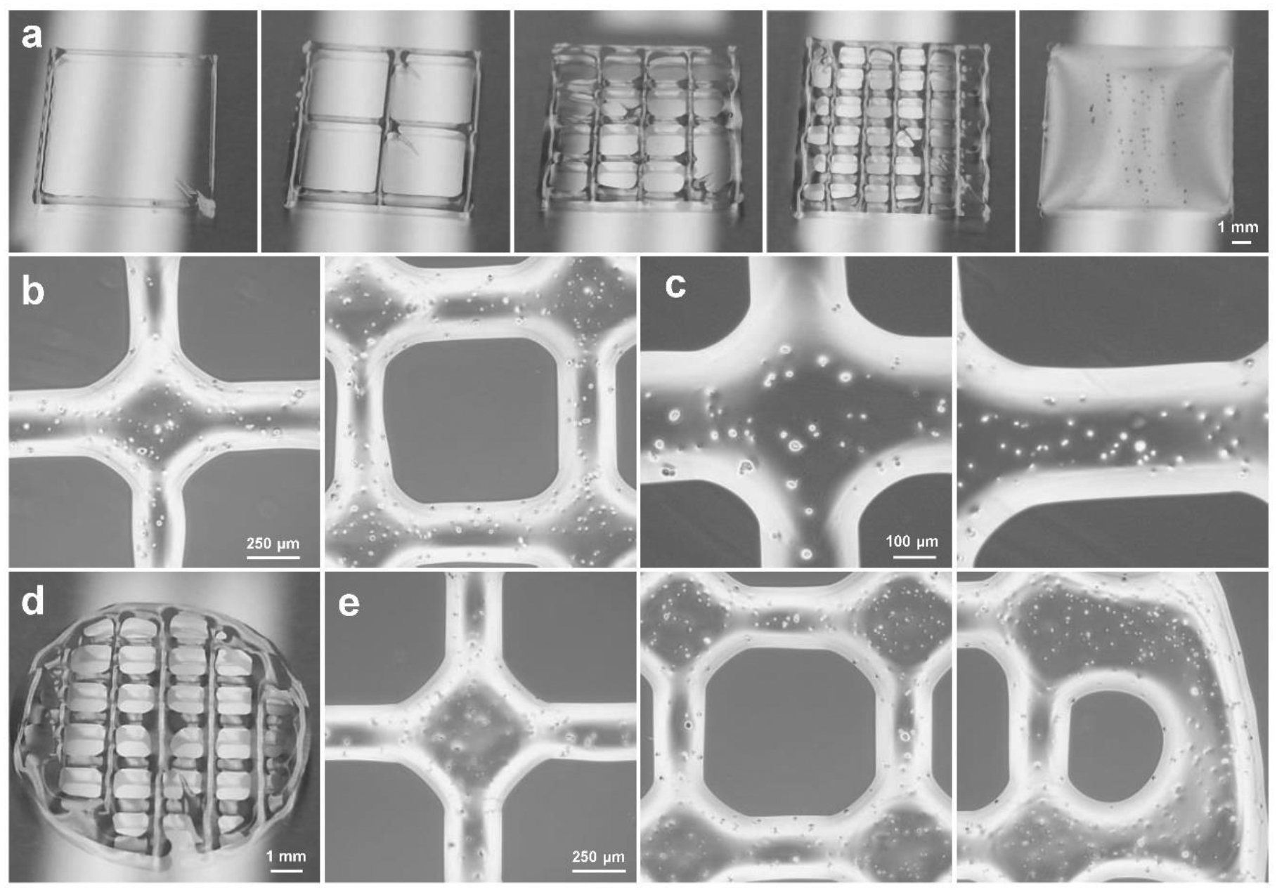
Fig. 4.

Bioprinted scaffolds morphology. a) Rectangular scaffolds printed with different infill densities. b) Microscopic images of the same printed scaffolds at different positions of the scaffolds and c) enlarged microscopic images of the scaffolds containing cells (1 × 10⁶ cells/mL of bioink). d) Photographs of the bioprinted circular scaffolds. e) Microscopic images at different locations along the bioprinted circular scaffolds.
