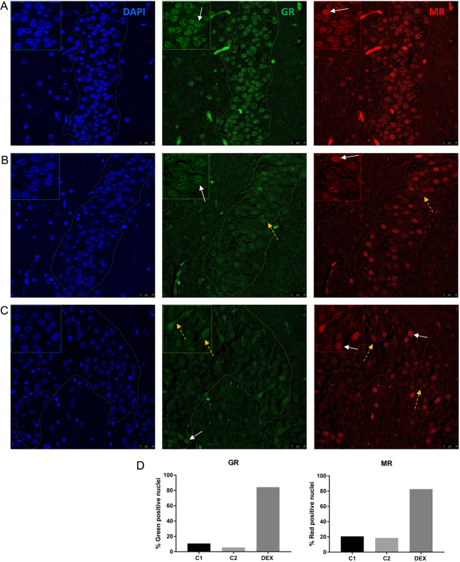
Figure 2.
Cell nuclear localization of GR and MR in human hippocampal dentate gyrus (DG) region. Immunofluorescence staining of cell nuclei (blue), GR (green) and MR with the 1D5 antibody (red) in (A) tissue of the dexamethasone-treated patient and in (B) tissue from the 5-year-old control patient and (C) tissue from the 11-year-old control patient. (D) The percentage of GR- and MR-positive cell nuclei in the different tissues. White arrows show nuclear MR staining and yellow dotted arrows show cytosolic MR staining. In the left corner, a magnification is shown, and the dotted line represents the region of interest.

 This work is licensed under a