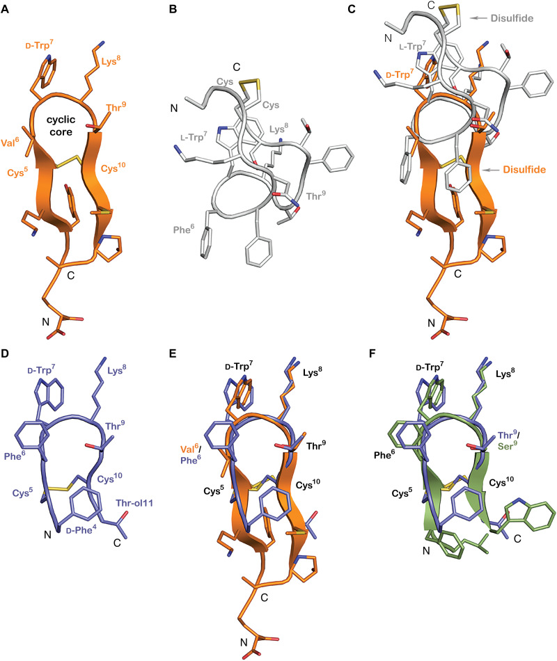
Fig. 4. Consomatin Ro1 and G1 are structural mimetics of the SS drug analog octreotide.
(A) X-ray structure of Consomatin Ro1 at 1.95-Å resolution (PDB ID: 7SMU). (B) Nuclear magnetic resonance (NMR) solution structure of human SS-14 obtained during heparin-induced fibril formation (PDB ID: 2MI1). (C) Alignment of the structure of Consomatin Ro1 (orange) with that of SS-14 (gray). (D) NMR solution structure of octreotide (PDB ID: 1SOC). (E) Alignment of the structure of Consomatin Ro1 (orange) with that of octreotide (purple) showing nearly identical backbone conformation and orientation of d-Trp7, Lys8, Thr9, and the disulfide bond, but differences in the amino acid composition and side-chain arrangements of Val/Phe6 and of residues outside the cyclic core. (F) Alignment of a homology model of Consomatin G1 (green), based on the structure of Ro1, with that of octreotide (purple), suggesting that the molecules share strong structural similarities. Numbering of residues according to that of Consomatin Ro1.

