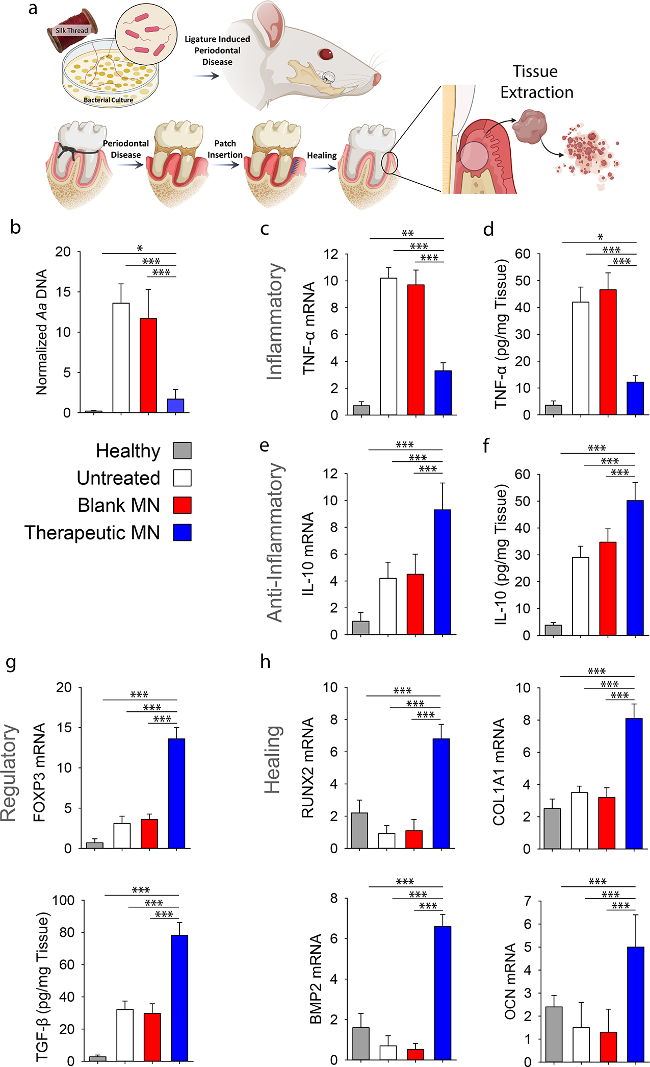
Figure 4. In vivo evaluation of the immunomodulatory effects in a rat model of periodontitis.

a, Ligature induced periodontal disease model in rats was used. a, Schematic representation of the proposed disease induction strategy patch placement. Mucoperiosteal flaps were elevated, uncovering the alveolar bone adjacent to the lingual aspect of the first maxillary molar to place MN at the defect site. At 8-weeks post MN insertion, buccal and palatal tissues of maxillary molars were isolated and dissociated to assess inflammatory status of tissue microenvironment. b, PCR analysis of A.a. bacterial ribosomal DNA in the periodontal tissue normalized by palatal tissue weight. c, Relative mRNA expression level of pro-inflammatory cytokine TNF-α. See also Figure S10. d, ELISA analysis of TNF-α level in the tissue. e, Relative mRNA expression level of anti-inflammatory cytokine IL-10. f, ELISA analysis of IL-10 in the tissue. g, Relative mRNA expression of Treg marker FOXP3 and ELISA analysis of TGF-β secretion. h, Relative mRNA expression level pro-healing genes RUNX2, COL1A1, BMP2 and OCN in the periodontal tissue. Healthy: healthy rats; Untreated: untreated periodontitis; Blank MN: MN patches without any therapeutic cargo; Therapeutic MN: MN patches containing both tetracycline- and IL-4/TGF-β-loaded SiMPs (Tetracycline: 0.5 mg; IL-2: 100 ng, IL-4: 40 ng, and TGF-β: 40 ng per patch). The presented data are expressed as mean ± SD (n= 5). The results were analyzed by using one-way ANOVA with post-hoc analysis. Statistical significance is indicated by * p < 0.05, ** p < 0.01 and *** p < 0.001.
