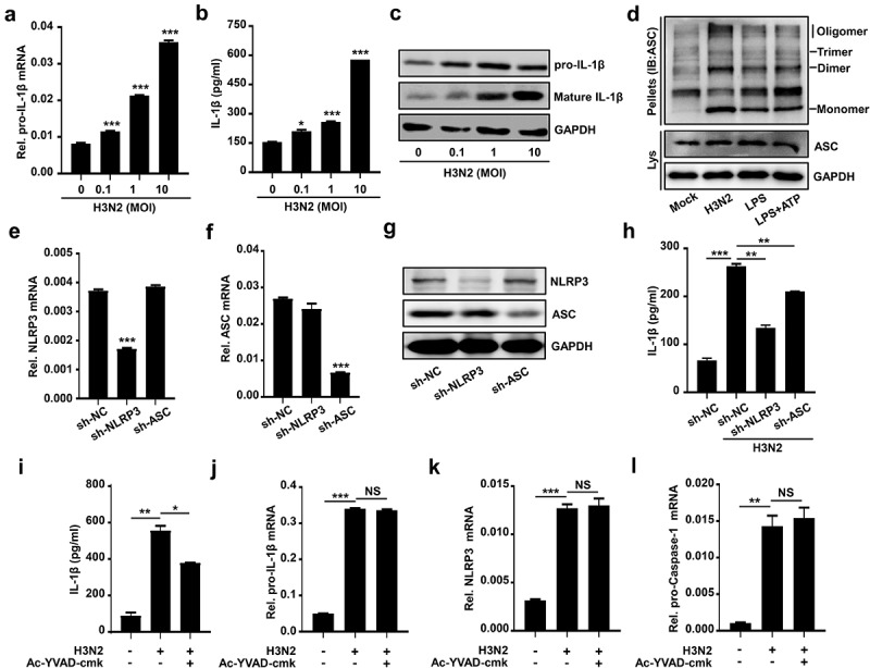
Figure 2.

IAV infection promotes NLRP3 inflammasome activation. (a–c) H3N2 (MOI = 0, 0.1, 1.0, and 10) infected THP-1 macrophages for 24 h. the pro-IL-1β mRNA level was analyzed by qPCR (a). Secreted IL-1β was detected by ELISA (b). the precursor and maturation of IL-1β, coupled with inter control GAPDH, were analyzed by using indicated antibodies by Western blotting (c). (d) THP-1 macrophages were treated with live H3N2 (MOI = 2), LPS (1 μg/ml) for 6 h, or LPS (1 μg/ml) for 6 h with ATP (5 mM) for 30 min, respectively. Lysates and ASC oligomerization were detected by indicated antibodies. (e–g) NLRP3 (e) and ASC (f) mRnas were assured by qPCR and NLRP3 (g), while ASC (g) proteins were detected by Western blotting in macrophages infected by lentiviruses expressing the indicated shRNA. (h) H3N2 (MOI = 2) infected macrophages stably expressing shRNA against target genes. IL-1β protein were detected by ELISA. (i–l) H3N2 (MOI = 2) infected macrophages for 24 h, and then the cells were treated with Ac-YVAD-cmk (1 mm) for 6 h. Secreted IL-1β was detected by ELISA (i). the mRNA levels of the indicated genes (i–k) were analyzed by qPCR.
