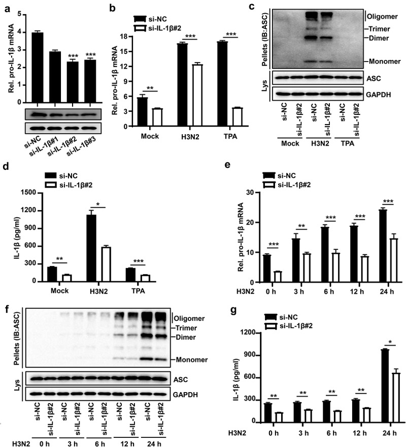
Figure 3.

IAV-Induced pro-IL-1β mRNA facilitates NLRP3 inflammasome activation. (a) A certain amount (50 nM) of si-NC (negative control), si-IL-1β#1, si-IL-1β#2, and si-IL-1β#3 were delivered into THP-1 macrophages by Lipofectamine 2000 for 36 h. the pro-IL-1β mRNA level was analyzed by qPCR and pro-IL-1β protein was detected by Western blotting. (b–d) the transfected macrophages were infected with H3N2 (MOI = 4) for another 24 h or were treated with TPA (50 ng/ml) for 6 h. the mRNA levels of pro-IL-1β (b) were quantified by qPCR. Lysates and ASC oligomerization were detected by the indicated antibodies (c). Secreted IL-1β was analyzed by ELISA assay (d). (e–g) A certain amount (50 nM) of si-IL-1β#2 or si-NC were delivered into macrophages, and then the cells were infected with H3N2 (MOI = 4) for different periods (0 h, 3 h, 6 h, 12 h, or 24 h). the mRNA levels of pro-IL-1β (e) were analyzed by qPCR. Lysates and ASC oligomerization were detected by the indicated antibodies (f). Secreted IL-1β was detected by ELISA (G).
