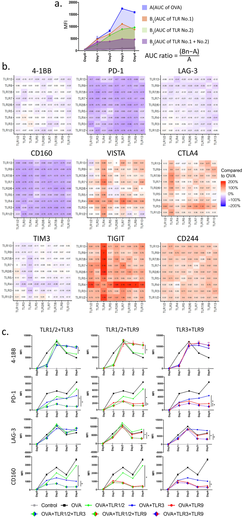
Figure 1.

Combinations of TLR agonists at the time of T-cell activation in vitro affect expression of T-cell checkpoint receptors: Splenocytes were prepared from the spleens of OT-1 mice and stimulated in vitro with the high-affinity SIINFEKL (OVA) peptide in the presence or absence of TLR agonists [TLR 1/2 (Pam3CSK4), TLR 3 (Poly I:C), TLR 4 (MPLAs), TLR 5 (FLA-ST), TLR 2/6 (FSL-1), TLR 7 (Gardiquimod), TLR 7/8 (R848), TLR 9 (ODN1826), or TLR 13 (ORN Sa19)] or their pairwise combinations. The median fluorescence intensity (MFI) of 4–1BB and T-cell checkpoint receptor expression on CD8+ T-cells were determined by flow cytometry, collected daily for 4 days, and computed as Area Under the Curve (AUC) using the trapezoid rule. (a) Calculation of AUC ratio to compare AUC of each receptor expression for each pairwise combination with that obtained following OVA stimulation alone, without TLR activation. (b) Heat-map demonstrating AUC ratio of each pairwise combination for 4–1BB and multiple T-cell checkpoint receptors. (c) Representative MFI plots showing 4–1BB, PD-1, LAG-3, and CD160 expression with the combinations of TLR1/2, TLR3, and TLR9 agonists. Results are from one experiment, with samples assessed in triplicate, and are representative of three similar, independent experiments. Asterisks represent significant (p < .05) differences as assessed by ANOVA.
