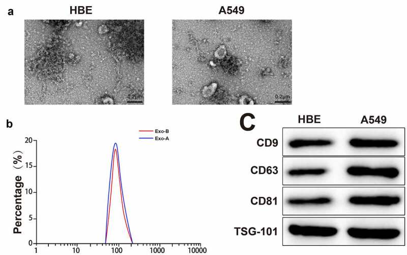
Figure 1.

Extraction and identification of LC-derived Exos.
Notes: The morphological characteristic of Exos was observed by TEM (A) (n = 3). The particle size of Exos was analyzed by NanoS90 (B) (n = 3). The expressions of marker proteins of Exos were measured by Western blot (C) (n = 3); ***P < 0.001; LC, lung cancer; Exos, exosomes; TEM, transmission electron microscope.
