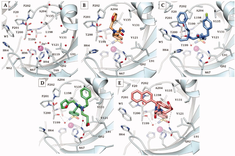
Figure 4.
X-ray crystallography: active site view of hCA IX-mimic in adduct with A) no inhibitor, B) 41, C) 42, D) 46, and E) 48. H-bonds are depicted as black dashed lines. Water molecules involved in water-bridged H-bonds are shown as red spheres. Amino acids are labelled with one letter symbols: D, Asp; E, Glu; F, Phe; H, His; I, Ile; L, Leu; N, Asn; P, Pro; Q, Gln; T, Thr; V, Val; W, Trp.

