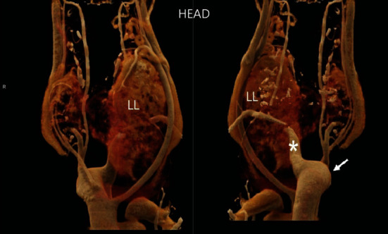
Figure 1.

CT scan three-dimensional reconstruction. Frontal and posterior views of the cervical region, showing the volumetric increase in the thyroid, pronounced on the left lobe (LL), and evidencing the presence of the aortic arch on the right side (arrow), with left subclavian artery with retroesophageal path (asterisk).
