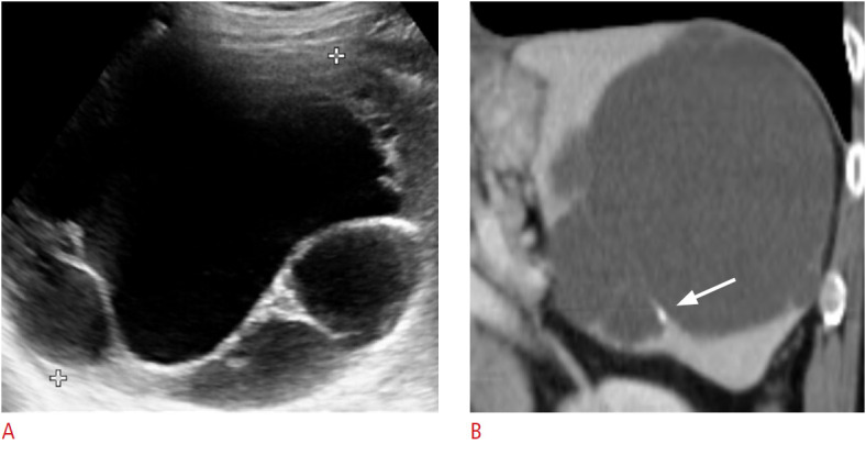
Fig. 12. A 74-year-old woman with splenic lymphangioma.

A. Transverse ultrasonography of the spleen shows an incidentally observed multiloculated cystic lesion measuring approximately 12 cm in the splenic upper pole. B. Coronal contrast-enhanced computed tomography image shows a multiloculated large cystic lesion with thin-wall calcification (arrow).
