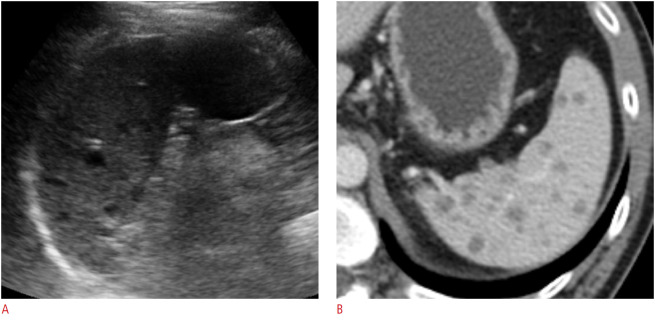
Fig. 7. A 35-year-old human immunodeficiency virus–positive man with miliary tuberculosis infection of the spleen.
A. Longitudinal ultrasonography (US) of the spleen shows multiple small (<1 cm) hypoechoic lesions in the splenic parenchyma. US-guided biopsy confirmed the presence of Mycobacterium tuberculosis. B. Axial contrast enhanced computed tomography image shows numerous, subcentimeter, hypodense nodular lesions throughout the spleen.

