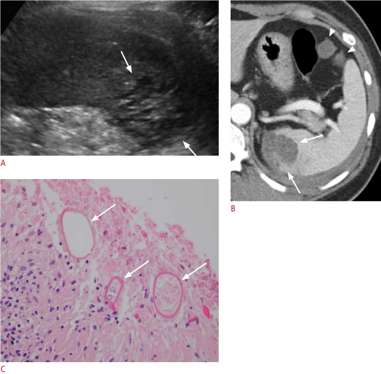
Fig. 8. A 44-year-old woman with paragonimiasis.

A. Longitudinal ultrasonography (US) of the spleen shows an approximately 3.5-cm clustered multicystic lesion (arrows). US-guided biopsy confirmed eggs from Paragonimus westermani. B. Axial contrast-enhanced computed tomography image shows a lobulated hypodense splenic lesion with clustered multiple cysts (arrows). Two small peritoneal cystic lesions (arrowheads) and left pleural effusion were also observed. C. Microscopic image with hematoxylin and eosin staining (×400) shows the ovoid parasite eggs with a thick shell (arrows) in necrotic splenic tissue, morphologically consistent with P. westermani.
