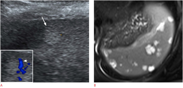
Fig. 9. A 33-year-old woman with a splenic hemangioma.
A. Longitudinal ultrasonography (US) of the spleen shows an incidentally found, discrete, round, echogenic nodule (arrow). Color Doppler US (box) showed peripheral vascularity (arrowhead) of the nodule. B. Axial magnetic resonance imaging; a T2-weighted image shows multiple well-defined, round, T2 high-signal-intensity lesions suggestive of hemangiomas.

