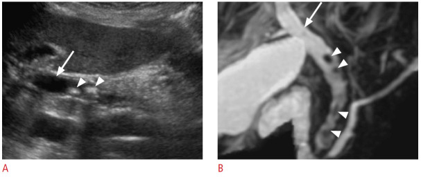
Fig. 4. Choledocholithiasis: a 91-year-old woman with abdominal pain and abnormal liver function tests.
A. Grayscale ultrasonography of the liver demonstrates a dilated common bile duct (arrow). There are several common bile duct stones (arrowheads) with posterior shadowing. B. Coronal magnetic resonance cholangiopancreatography shows biliary ductal dilatation (arrow) and stones (arrowheads) throughout the common bile duct.

