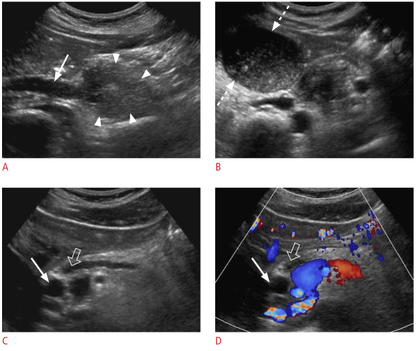
Fig. 9. Periampullary tumor: a 41-year-old man with a 2-week history of epigastric pain, nausea, and mildly elevated liver enzymes and lipase.
Grayscale (A-C) and color Doppler (D) ultrasonography are shown. A. There is a large, heterogeneous, hypoechoic mass lesion (arrowheads) at the head of the pancreas causing bile duct dilatation (arrow). B. The gallbladder is filled with sludge and distended (dashed arrows). C, D. Simultaneous dilatation of the common bile duct (arrows) and pancreatic duct (open arrows) due to the pancreatic head mass (double duct sign) is shown. The pathology was neuroendocrine tumor.

