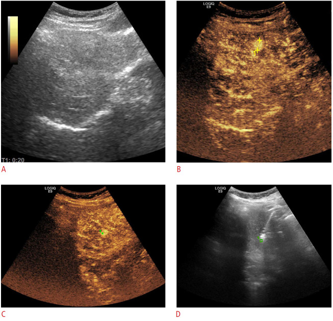
Fig. 1. Hepatocellular carcinoma.
A. Grayscale ultrasonography could not detect a small recurrent hepatocellular carcinoma nodule (11 mm), previously depicted on a computed tomography scan. B, C. During contrast-enhanced ultrasonography, the hyperenhancing nodule became apparent (B, marked in yellow), and allowed GPS marking (C, green dot). D. Subsequently, microwave ablation was performed in grayscale mode, targeting the previously marked area.

