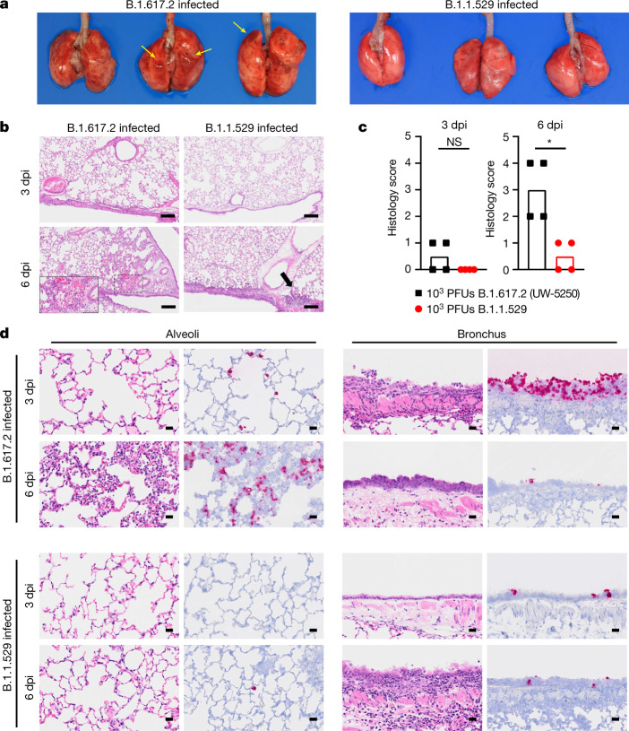
Fig. 3. Pathological findings in the lungs of SARS-CoV-2-infected Syrian hamsters.
Hamsters were inoculated with 103 PFU of B.1.617.2 or B.1.1.529 and euthanized at 3 and 6 dpi (n = 4). a, Macroscopic images of the lungs obtained at 6 dpi. Yellow arrows indicate haemorrhage. b, Lung sections from animals infected with B.1.617.2 or B.1.1.529. Scale bars, 200 µm. Focal alveolar haemorrhage in B.1.617.2-infected animals at 6 dpi is outlined and shown at higher magnification in the inset (scale bar, 100 µm). Black arrow indicates focal inflammation. c, Histopathological score of pneumonia based on the percentage of alveolitis in a given section using the following scoring: 0, no pathological change; 1, affected area (≤10%); 2, affected area (<50%, >10%); 3, affected area (≥50%); an additional point was added when pulmonary edema and/or alveolar haemorrhage was observed. Data are median score (n = 4; *P = 0.0286; two-tailed Mann–Whitney test). d, RNA in situ hybridization for SARS-CoV-2 viral RNA. Representative images for the alveoli and bronchi of hamsters infected with B.1.617.2 or B.1.1.529 (n = 4) virus at 3 or 6 dpi are shown. Scale bars, 20 µm. See Supplementary Table 1 for more information.

