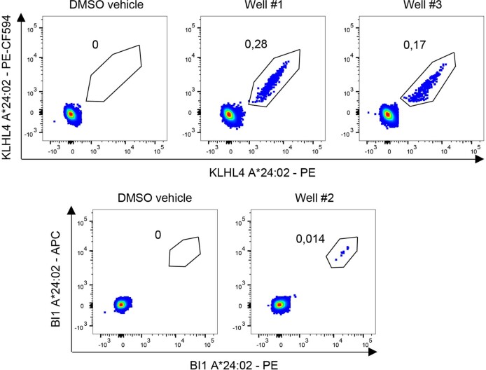
Extended Data Fig. 7. T cell reactivity against two substitutant peptides.
Flow cytometric analysis of CD8+ T cells following co-culture of naive CD8+ T cells and autologous monocyte-derived dendritic cells pulsed with peptide or DMSO vehicle. Plots show T cells reactive to SA-phycoerythrin (PE) and SA-phycoerythrin-CF594 (PE-CF594)-labelled pMHC multimers complexed with the KLH4L substitutant peptide YFDPHTNKF (Wells #1 and 3), or reactive to SA-PE and SA-allophycocyanin (SA-APC)-labelled pMHC multimers complexed with the BI1 (TMBIM6) substitutant peptide EHGDQDYIF (Well #2).

