Figure 3.
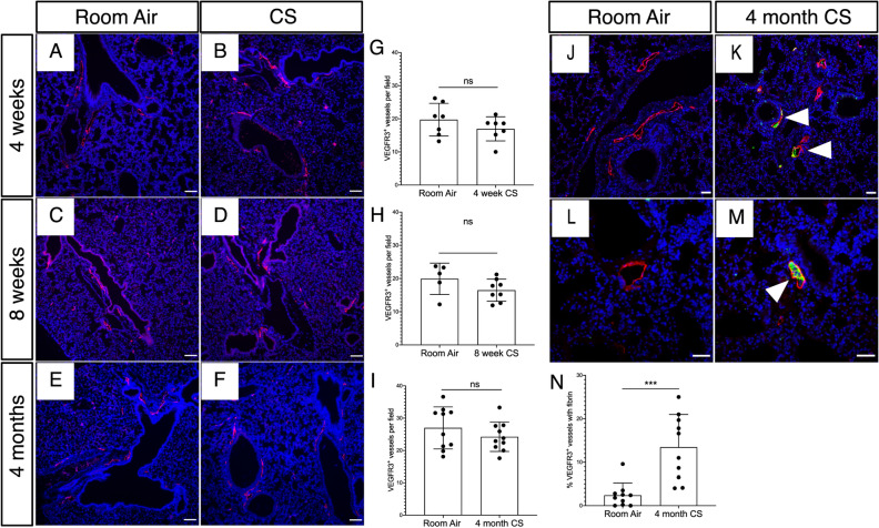
Lymphatic thrombosis after CS exposure in mice. Mice were exposed to full body CS and the lungs were harvested for fluorescent immunohistochemical analysis. (A–F) Representative images of immunohistochemical staining of lung tissue for the mouse lymphatic marker VEGFR3 (red) in mice exposed to CS for 4 weeks, 8 weeks, and 4 months compared to identically housed age matched mice exposed to room air. (G–I) Quantification of lung lymphatic vessel density in CS-exposed and control mice at the indicated time points. (J–M) Analysis of lung lymphatic thrombosis using immunohistochemical staining for VEGFR3+ (red) and fibrinogen (green). Thrombosed lymphatics indicated with arrowheads. (N) Quantification of lymphatic thrombosis, as expressed by percentage of VEGFR3+ vessels with luminal fibrin after 4 months of CS exposure. Quantification lung lymphatic vessels was performed by counting VEGFR3+ lymphatics in 10 × images of lung tissue sections. At least 5 10 × images were used for each tissue sample and the average lymphatic number was determined. Lymphatic thrombosis was quantified as the percentage of VEGFR3+ lung lymphatics with luminal fibrin in each 10 × image and was averaged from at least 3 10 × images. ***P < 0.001. ns not significant. Scale bars in (A–F,J,K) = 25 μm. Scale bars in (L,M) = 50 μm.

