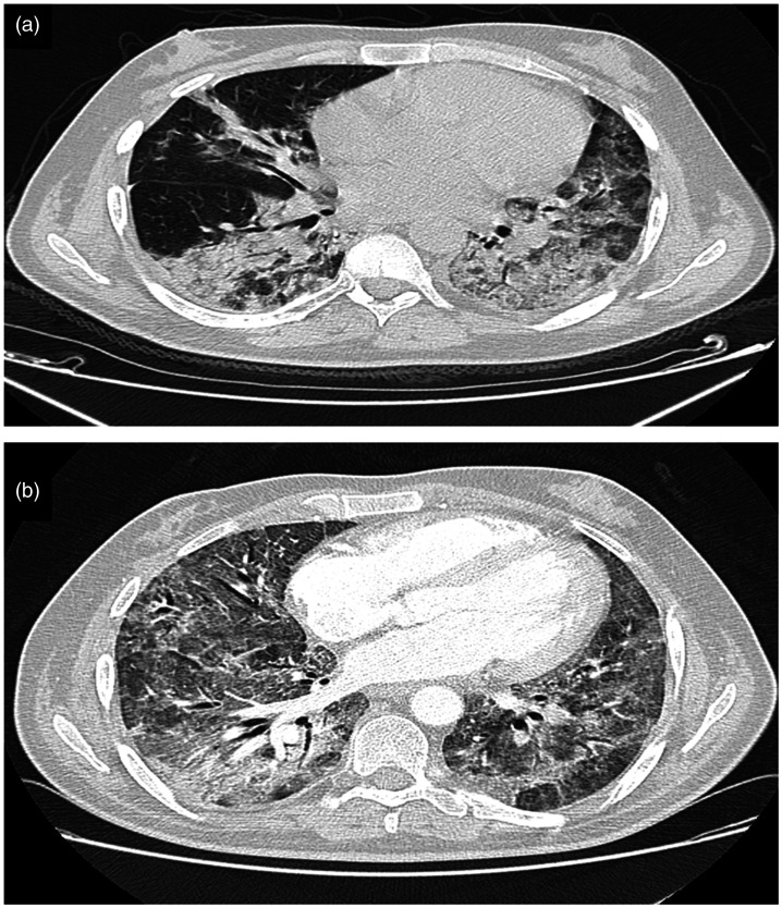
Figure 1.
(a) Axial nonenhanced chest computed tomography image (lung window) showing bilateral ground-glass opacities typical of SARS-CoV-19 infection with pulmonary involvement estimated between 25% and 50%. (b) Axial contrast-enhanced chest computed tomography image (lung window) showing worsening of the lesions with estimated pulmonary involvement of >75%.

