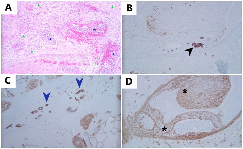
Figure 2.
Pulmonary tumor thrombotic microangiopathy: (A): H&E, 10×: H&E-stained slide showing fibrocellular intimal proliferation with spindle-shaped cells (green asterisks) surrounding the pulmonary tumor emboli (blue asterisks) and completely filling the lumen of this vessel, (B) Immunostaining for CK-7, 10×: CK-7 highlights tumor cells (black arrowhead) within the lumen of muscular arteries, adjacent to intimal proliferation, (C) Immunostaining for CK-7, 10×: CK-7-positive tumor cells (blue arrowheads) present in lymphatics, and (D) Immunostaining for SMA, 10×: smooth muscle actin (SMA) highlights proliferation of smooth muscle cells within tunica intima (intimal proliferation) (black asterisks).

