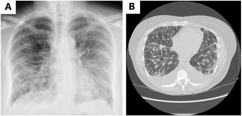
Figure 3.
(A) Chest radiograph at initial presentation revealing diffuse reticular opacities and tiny bilateral pleural effusions and (B) CT scan of chest without contrast revealing bilateral pleural effusions with diffuse interstitial septal thickening and scattered ground-glass opacities.
Abbreviation: CT, computed tomography.

