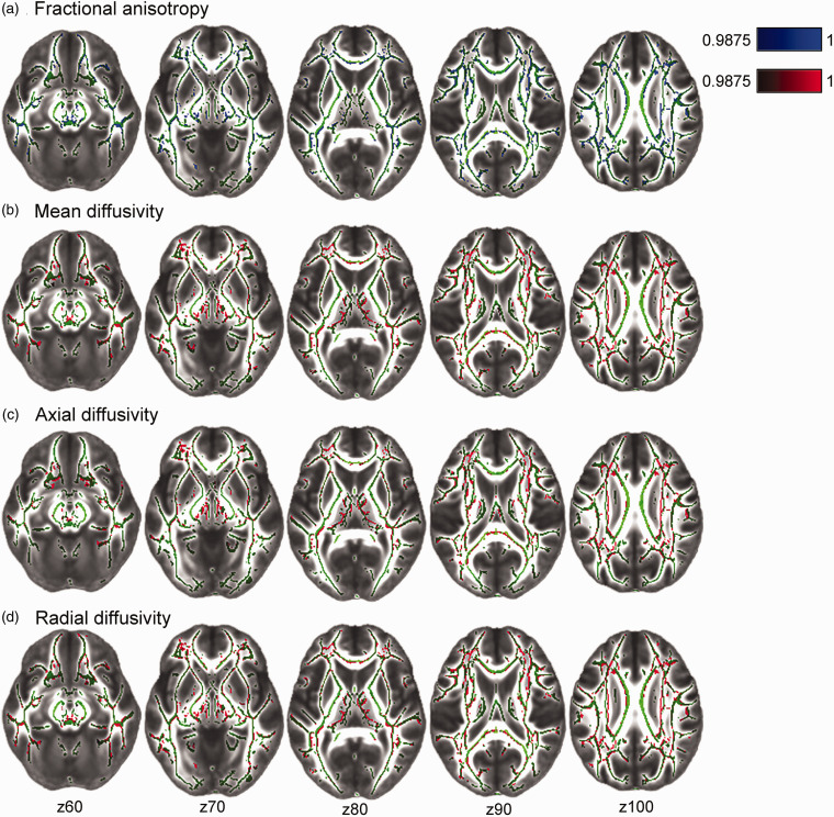
Figure 3.
Voxel-wise associations between cerebral small vessel disease (SVD) burden and diffusion tensor imaging measures: fractional anisotropy (FA; A) mean diffusivity (MD; B); axial diffusivity (AD; C), and radial diffusivity (RD; D). Statistical maps were overlaid on the FMRIB58_FA standard image. Green tracts depict the standardized mean FA skeleton. Higher SVD burden is associated with lower FA (in blue), and higher MD, AD, RD (in red). All images are TFCE-corrected statistics thresholded at 1-p values of 0.9875, i.e. representing Bonferroni-corrected p < 0.0125 to adjust for multiple comparisons across 4 DTI metrics. Images are also FWE-corrected for multiple voxel-wise comparisons. Analyses were adjusted for age, sex, education, MRI scanner model, antihypertensive use, body mass index, and mean arterial pressure measured at the time of the MRI scan.

