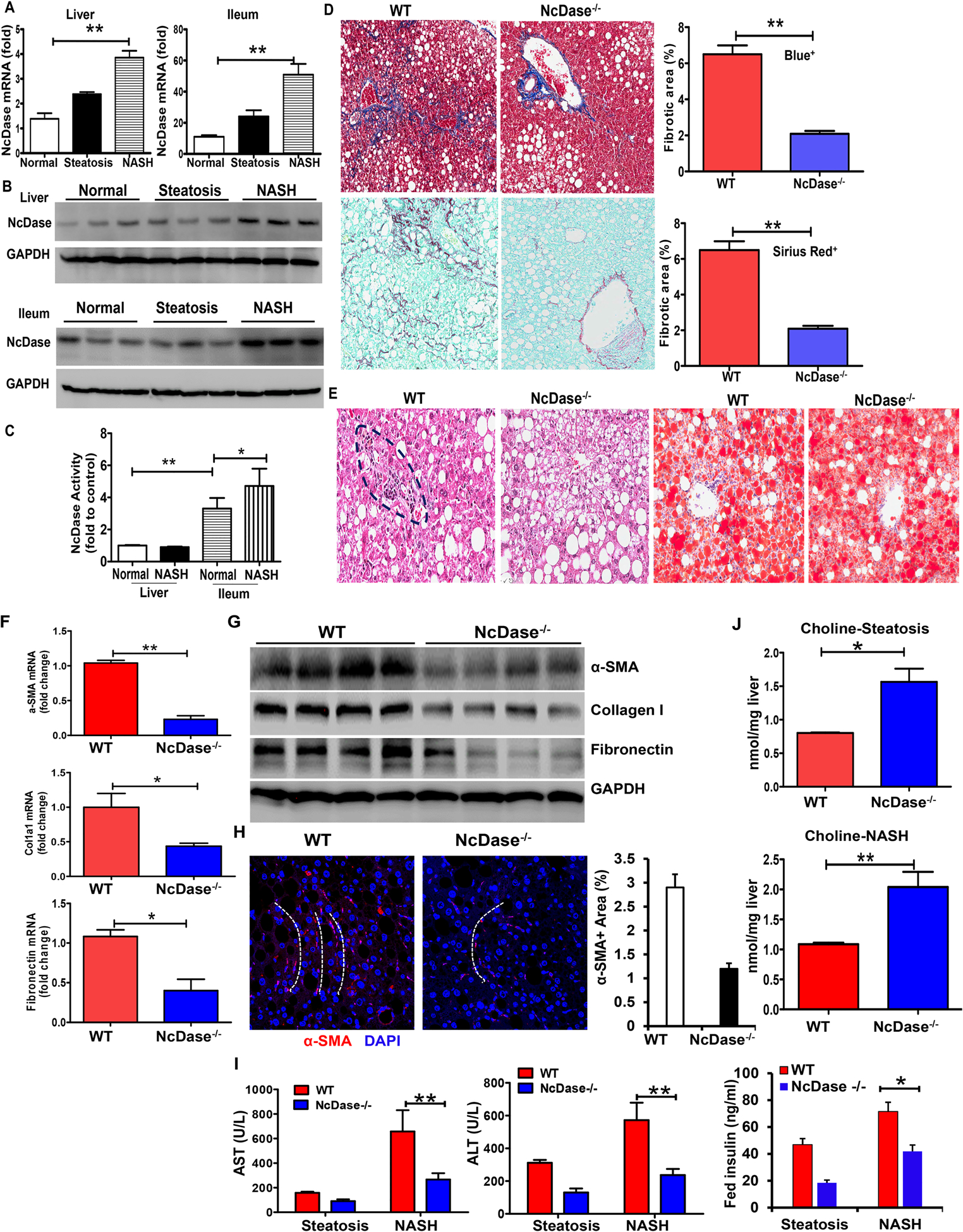
Figure 1. NcDase deficiency reduces liver inflammation and fibrosis in HFD-fed mice.

WT mice or NcDase−/− mice were fed the HFD diet for 7 months (inducing steatosis) or 14 months (inducing NASH).
(A) Quantification of NcDase mRNA in liver and ileum in normal, steatotic and NASH mice.
(B) Immunoblots of NcDase in liver and ileum in normal, steatotic and NASH mice.
(C) The activity of NcDase in liver and ileum in normal and NASH mice
(D-H) Liver section from mice with induction of NASH.
(D) Staining for Masson’s trichrome and Aniline blue-positive area (Top) and for Sirius red and Sirius red-positive area (Bottom).
(E) Staining for H&E and Oil red O.
(F) mRNA levels of the indicated genes related to fibrosis in liver.
(G) Immunoblot of the protein related to fibrosis in liver.
(H) α-SMA immunofluorescence (red) and DAPI counterstain (blue) and quantification. The white dot lines indicate the fibrotic pattern of α-SMA+(red) cells.
(I-J) Serum ALT and AST, and insulin levels (I) or liver choline levels (J) in mice with steatosis or NASH.
Mean ± SEM; n = 10, *p < 0.05, **p < 0.01. Abbreviation: GAPDH, glyceraldehyde 3-phosphate dehydrogenase.
